The 1.4 eV nickel color centers in diamonds
Photoluminescence study at low temperatures (80–300 K)
Abstract
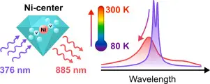
Diamonds synthesized at high pressures and temperatures (HPHT) often contain defects associated with nickel impurities. We present a detailed spectroscopic study of the 1.4 eV nickel-related color centers in microdiamonds over the temperature range of 79–298 K. The temperature dependences of the intensity, linewidth, zero-phonon transition energy and photoluminescence lifetime are obtained. Also temperature dependences of the full spectrum of the defect with a phonon band up to 1 eV and the Debye–Waller factor are analyzed. The results show that the width of the zero-phonon line varies as T^3 with temperature, and the spectral peak undergoes a blueshift by up to 3.7 meV. These findings provide a more comprehensive understanding of the luminescence properties of Ni centers in diamond, which have promising potential for all-optical thermometry in the near-infrared range.
Introduction
Diamond has long been considered an exceptional material due to its unique mechanical, thermal and optical properties [1]. As a wide-band semiconductor, diamond crystals contain numerous luminescent defects, known as color centers, that emit light from the ultraviolet (UV) to near-infrared (near-IR) range [2]. Although several hundred color centers are identified in natural and synthetic diamonds, only a few of them have been extensively studied [2], [3]. The nitrogen vacancy (NV) defect is the most well-known of all types of centers. Also, in the last decade, there has been a growing interest in vacancies of group IV elements, such as silicon (SiV), germanium (GeV), tin (SnV), and lead (PbV), due to their narrow zero-phonon lines (ZPLs) and high () Debye–Waller factors [4], [5]. Diamond crystals with color centers have become a promising material for a variety of new applications, including biovisualization [6], [7], electric and magnetic field sensing [8], [9], [10] and thermometry on the micro- and nanoscale [11]. In addition, diamond color centers exhibit stable single-photon emission at room temperature [12], [13], [14], [15], making them potential candidates for future architectures of quantum information processing and quantum communications [16], [17], as well as for integrated photonic systems [1].
Optical (or luminescent) thermometry is one of the potential applications of diamond defects [18]. Luminescent thermometry is a promising technique that allows temperature detection remotely. This method is useful in applications where conventional contact sensors are ineffective, such as with fast-moving objects, in harsh environments or in strong electromagnetic fields [19]. Additionally, it allows for real-time measurement of temperature in micro- and nanoscale systems [20].
To date, optical thermometry has been demonstrated for various luminescent systems, including organic dyes and coordination compounds [21], [22], quantum dots [23], color centers in hexagonal boron nitride [24] and diamond [18]. Among these, diamond stands out due to its advantages such as high photostability, biocompatibility, resistance to extreme environmental conditions and the ability to operate over a wide range of temperatures [25]. Diamond-based thermometric methods can be divided into spin-based and all-optical thermometry. The spin-based method relies on a shift in the spectrum of optically detected magnetic resonance (ODMR) signal from NV centers. However, due to the technical complexity of the implementation, the scope of this method is limited [18]. The second method is more accessible and uses the temperature dependence of the shape, intensity of the spectrum, ZPL position or luminescence lifetime. All-optical thermometry demonstrated in 2010 for NV centers [26] has since been shown for vacancies of group IV elements [27], [28], [29], the IR defect of the silicon complex [30] and Ni-based centers in diamond [20], [31], [32], [33], [34].
Nickel is often incorporated into the crystal structure of synthetic diamonds grown using the HPHT method. This method is widely used on an industrial scale. The most well-known Ni defects are centers with ZPLs at 1.4 eV (885 nm), 2.56 eV (484 nm) and 3.1 eV (400 nm) [35]. Depending on the doping method, nickel can form many other diamond defects. Recently, diamonds containing nickel–nitrogen complexes with ZPLs at 1.56 eV and 1.66 eV were obtained in the HPHT process with ongoing spontaneous crystallization (without the use of seed microcrystals). For these diamonds the temperature dependence of their photoluminescence characteristics was studied [36]. Previously, the NE8 nickel–nitrogen complex with ZPL at 796 nm and a nickel-silicon complex with ZPL at 768 nm were produced using chemical vapor deposition (CVD) method. In addition, single-photon generation was demonstrated for these centers [37]. Near-infrared emitters are especially promising in biological applications, as tissues have weak absorption and luminescence in the 650–1350 nm wavelength range. Among these emitters the 1.4 eV Ni center has an emission band lying approximately in the middle of the biological transparency window, making it promising for biovisualization, biomarker applications [38], [39] and for near-infrared thermometry [32].
The 1.4 eV centers observed in both synthetic and some natural diamonds have been studied using electron paramagnetic resonance, cathodoluminescence, and optical spectroscopy [38]. Despite this, there has been no clear understanding of the defect structure for several decades. It has been suggested that this center is associated with a positively charged Ni defect in the interstitial (Ni) [40] or substitutional position (Ni) [39]. Recently, a theoretical group using the ab initio method of magnetic spectroscopy has shown that the 1.4 eV center consists of a negatively charged Ni atom in a split-vacancy configuration (NiV) [41]. This configuration is similar to the group IV vacancies, for which temperature dependences of linewidths and ZPL positions over a wide temperature range have been obtained [4]. These dependences are important for understanding the mechanisms involved in electronic transitions and for thermometry applications.
As far as we know, temperature spectral studies of 1.4 eV Ni centers were conducted near room temperature or in a limited range between helium and nitrogen temperatures [34], [39], [40]. Thus, in this study we investigate the spectra and photoluminescence lifetimes of 1.4 eV Ni centers in microdiamonds for the temperature range from 79 K to 298 K (−194 °C to 25 °C). The temperature dependences of the ZPL linewidth, its position, intensity and luminescence lifetime were obtained. It is shown that the linewidth broadens according to the T^3 law, while the ZPL peak blueshifts with increasing temperature.
Materials
Commercial diamond microcrystals (Mono-&Polycrystalline Diamond Powder, China) of type Ib synthesized by the HPHT method and milled by the manufacturer were used in the study. These diamonds are synthesized using nickel and other catalysts. The sample was a monodisperse powder of microdiamonds with an average size of 30 μm. Fig. 1 shows images of microcrystals deposited on glass under an optical and scanning electron microscope (SEM). The crystals have a different shape with multiple small
Results
Fig. 2a shows the PL spectra of the microdiamond sample at room temperature (298 K) and liquid nitrogen temperature (79 K). The spectra were recorded on a visible-range spectrofluorometer. The excitation was performed with a 376 nm laser at a power of 1.5 mW. As can be seen from Fig. 2a, there are several spectral bands in diamond corresponding to different color centers. At low temperatures, we can clearly see the doublet at 484 nm (2.56 eV) and 488 nm (2.54 eV) associated with the negative
Discussion
According to the recent calculations within the spin-polarized density functional theory (DFT), the 1.4 eV center is a NiV defect [41], [47]. It has a split-vacancy configuration, i.e. the Ni atom is located between two carbon vacancies, forming a bond with Dsymmetry (see the inset in Fig. 3). This configuration is also typical for group IV diamond defects and was studied in detail previously [4], [48]. Nevertheless, the 3d orbitals of the Ni atom play an additional role in the energy
Conclusion
In conclusion, we have investigated the main photoluminescence parameters of the 1.4 eV Ni centers in microdiamonds, such as spectra and lifetimes for a wide temperature range from 79 K to 298 K. Temperature dependences of the ZPL width, its position and intensity were obtained. It was shown that the line broadening follows the T3 law with increasing temperature. Also results demonstrates the anomalous blue shift of the ZPL position in the range from 79 K to 200 K that depends on temperature.
https://www.sciencedirect.com/science/article/abs/pii/S0925346725006226